Next-generation 7T MRI scanner designed for ultra-high resolution human brain imaging
To increase granularity in human neuroimaging, we designed and built a next-generation 7 Tesla MRI scanner to reach ultra-high resolution by employing several advances in hardware. To improve spatial encoding, we developed a head-only asymmetric gradient coil "Impulse gradient" with unprecedented speed with an additional third layer of windings. To increase SNR, we integrated a multi-channel receiver systems, raising signal in the cerebral cortex and enabling high acceleration factors. A multi-channel transmit system reduced power deposition and improved image uniformity. The scanner routinely performs functional imaging studies at ultra-high 0.35 – 0.45 mm isotropic spatial resolution, to reveal cortical layer functional activity, achieves high angular resolution in diffusion imaging and reduces acquisition time in both functional and structural imaging.
Speaker
NMR in Photo- and Organocatalysis – Pushing the Limits
The detection and characterization of intermediates in catalytic reactions is crucial for the rational optimization of reaction conditions. However, in many rapidly expanding fields of asymmetric catalysis, mechanistic studies as well as structural investigations on intermediates or intermolecular interactions are scarce. In this talk I will present techniques and methods to extend the application of NMR in photocatalysis and ion pair catalysis and explain their impact on examples. First our LED based NMR illumi-nation device [1] will be introduced together with the new triple combination illumina-tion/NMR/UV [2] and an NMR access to intermediates below the detection limit [3]. These methods allow for new insights into one- versus two-electron processes usually inaccessible to UV/Vis [4], the inclusion of radical species into NMR reaction profiles [2], the structure elucidation of thermally labile photoswitches [2], the sequencing of tiny intermediates [5], and even insights into photosteps in photocatalysis [6]. Last, the structure elucidation, H-bond analysis, reactivity/selectivity understanding and experimental transition state combination analysis of chiral phosphoric acids in ion pair catalysis is demonstrated [7]. Here, the NMR time scale could be extended so far that even the switching of a single hydrogen bond can be detected experimentally [8]. The translation of structural insights to synthetic applications is shown at two examples: Higher stereoselectivities were obtained for a whole bundle of ion pair catalysts combining ground- and transition state analysis with dispersion donor effects [9]. Furthermore, catalyst ion pair analysis of reaction intermediates revealed a dimeric pathway with inverse stereoselectivity so far hidden in ion pair catalysis [10].
[1] C. Feldmeier, H. Bartling, E. Riedle, R.M. Gschwind, J. Magn. Res., 2013, 232, 39.
[2] A. Seegerer, P. Nitschke, R.M. Gschwind, Angew. Chem. Int. Ed. 2018, 57, 7493.
[3] L. Nanjundappa, A. Seegerer, J. Hioe, R.M. Gschwind, JACS 2018, 140, 1855.
[4] C. Feldmeier, H. Bartling, K. Magerl, R.M. Gschwind, Angew. Chem. Int. Ed., 2015, 54, 1347.
[5]. S. Wang, L. Nanjundappa, J. Hioe, R. M. Gschwind, B. König, Chem. Sci., 2019, 10, 4580. [6] N. Berg, S. Bergwinkl, P. Nuernberger, D. Horinek, R.M. Gschwind, JACS 2021, 143, 724. [7] JACS, 2016, 138, 15965; JACS, 2016, 138, 16345; JACS 2017, 139, 6752; Chem. Sci. 2019, 10, 5226; Chem. Sci. 2019, 10, 10025; Chem. Sci. 2020, 11, 4381; Chem. Sci. 2021, 12, 15263; JACS, 2022, 144, 19861.
[8] L. Nanjundappa, J. Hioe, J. Gramueller, R. M. Gschwind, JACS 2019, 141, 16398.
[9] J. Gramüller, M. Franta, R. M. Gschwind, JACS 2022, 144, 1986.
[10] M. Franta, J. Gramüller, P. Dullinger, D. Horinek, Angew. Chem. Int. Ed. 2023, 62, e202301183.
Speaker

Profile
Heterogeneous Catalysts for Hydrogenative and Non-Hydrogenative Parahydrogen Induced Polarization
Hyperpolarization from parahydrogen (pH2),1 or PHIP, shows outstanding potential for clinical MRI signal enhancement.2 PHIP is a rapid, scalable, and inexpensive method that can achieve high hyperpolarization levels using either homogeneous or heterogeneous catalysis. Advantageously, heterogeneous catalysis over supported noble metals affords production of continuous streams of hyperpolarized molecules that are free of polarizing agents or catalyst residues. However, their performance is compromised by rapid randomization of parahydrogen spin order after dissociative chemisorption. Several catalyst design strategies have been employed for prolonging the lifetime of singlet order in H ad-atom pairs sourced from parahydrogen. In one approach, the H ad-atom diffusion on Pt surfaces was suppressed by introduction of Sn to form Pt-Sn intermetallic phases, resulting in significant improvement in hydrogenative PHIP hyperpolarization.3 Remarkably, one intermetallic phase, namely Pt3Sn, facilitated the observation of non-hydrogenative surface-mediated hyperpolarization of protons in water and other solvents.4 Norcot recently reported that similar signals can be observed by adding benzoquinone to a suspension of a commercial Pt/C catalyst at room temperature.5 These results will be discussed with an emphasis on the molecular mechanisms and spin dynamics.
(1) Bowers, C. R.; Weitekamp, D. P. J Am Chem Soc 1987, 109
(18), 5541–5542. doi.org/10.1021/ja00252a049.
(2) Hovener, J. B. et al. Angewandte Chemie International Edition 2018, 57
(35), 11140–11162. doi.org/10.1002/anie.201711842.
(3) Zhao, E. W. et al. Angewandte Chemie - International Edition 2017, 56
(14), 3925–3929. doi.org/10.1002/anie.201701314.
(4) Zhao, E. W. et al. Chem 2018, 4
(6), 1387–1403. doi.org/10.1016/j.chempr.2018.03.004.
(5) Norcott, P. L., J Am Chem Soc 2023, 145
(18), 9970–9975. doi.org/10.1021/JACS.3C01593
Speaker

Profile
EPR as a tool to understand the molecular basis of chlorite dismutases, a class of industrially relevant biocatalysts
Chlorite (ClO2-) and hypochlorite (ClO-) are strong oxidants commonly employed as bleaching agents or disinfectants. However, concerns have been raised about their presence in the environment as pollutants. Chlorite dismutases (Clds) are heme b-containing oxidoreductases which possess the unique ability to decompose ClO2- into harmless Cl- and O2. Investigations into the underlying reaction mechanism thus open ways for potential biotechnological and bioremediation applications [1]. This talk will focus on how the heme-pocket architecture impacts protein stability and binding of substrate (or substrate analogues) and highlight the role of the fully conserved arginine at the distal site of the heme moiety [2-4]. Furthermore, during the reaction of Clds with either ClO2- or ClO-, short-lived intermediates involving transient radicals are formed. Using a rapid freeze-quenching device in combination with electron paramagnetic resonance techniques, attempts are made to trap the fast-evolving species. The strengths, but also the pitfalls of using EPR techniques in the research of heme proteins will be highlighted [5].
1. S. Hofbauer et al., Biotechnology journal (2014) 9, 461-473.
2. D. Schmidt, I. Serra, et al., Biochemistry (2021) 60, 621–634.
3. I. Serra, D. Schmidt et al., Journal of Inorganic Biochemistry (2022), 227, 111689.
4. D. Schmidt, et al., Biochemistry (2023), 62, 835–850.
5. I. Serra, et al., Applied Magnetic Resonance (2022) 53, 1105-1119.
Speaker

Profile
Signs of a ‘Time Crystal’ in a Surprising Place
The periodic spatial arrangement of atoms in a crystal like NaCl has been known for a century. The concept of a "time crystal" is a much more recent development, just a decade old. In 2016, theorists predicted the existence of a Floquet/discrete time crystal (DTC), a prototypical example of a novel phase of matter that awaits discovery in many-body systems driven out of equilibrium. Thus, it was exciting when in 2017, independent experiments using ion traps and diamond NV centers both reported that they had found signatures of the DTC. Here, we present nuclear magnetic resonance (NMR) observations of DTC signatures in a third, strikingly different system: an ordered spatial crystal of ammonium dihydrogen phosphate (ADP). We will explain how we accidentally ended up probing a new corner of parameter space, and why our results appear to challenge earlier assumptions about the conditions needed for DTCs to form. Moreover, we will describe how we expanded the NMR toolbox to develop a novel DTC-echo, revealing the hidden coherence produced by the DTC sequence. The twists and turns that led to these serendipitous discoveries are well described by Louis Pasteur’s quote: “chance favors the prepared mind”.
References:
1. Jared Rovny, Robert L. Blum, and Sean E. Barrett, Phys. Rev. Lett. 120, 180603 (2018).
2. Jared Rovny, Robert L. Blum, and Sean E. Barrett, Phys. Rev. B 97, 184301 (2018).
Profile
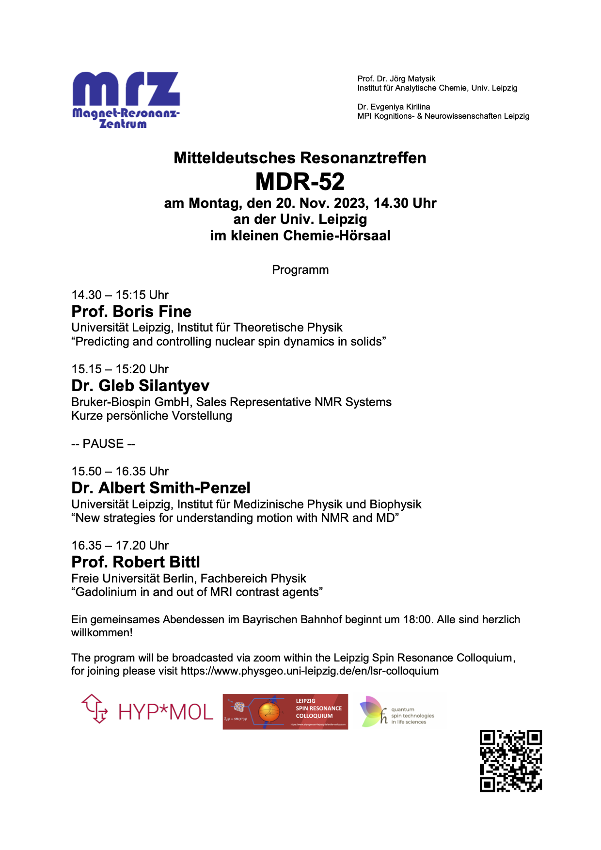
The Workshop program
Mitteldeutsches Resonanztreffen MDR-52
Exploring Tangible Cultural Heritage with Mobile NMR
Cultural heritage defines our identity. Tangible cultural heritage is treasured in museums, excavation sites, and nature parks. Their preservation and restoration strategies often rely on nondestructive methods of analysis. One such method is NMR known from spectroscopy for chemical analysis and imaging for medical diagnostics. Mobile NMR employs portable instruments which measure relaxation and diffusion parameters in stray magnetic fields from regions outside the magnet. The NMRMOUSE (Mobile Universal Surface Explorer) is a compact stray-field NMR sensor, which is being used to acquire depth profiles non-destructively through layered objects. How compact mobile NMR instruments work will be explained, and their use in analyzing objects of cultural heritage will be illustrated with measurements of antique Roman frescoes at excavation sites, paintings and master violins in museums, mummies and bones, and biofilms in Yellowstone National Park.
References:
1. B. Blümich, The Incredible Shrinking Scanner, Scientific American 299 (2008) 68
2. B. Blümich, S. Haber-Pohlmeier, W. Zia, Compact NMR, de Gruyter, Berlin, 2014
3. C. Rehorn, B. Blümich, Cultural heritage studies with mobile NMR, Angew. Chem. Int. Ed. 57 (2018) 7312
4. B. Blümich, Concepts and Applications of the NMR-MOUSE, In: D.M. Bastidas, E. Cano, eds., Advanced Characterization Techniques, Diagnostic Tools and Evaluation Methods in Heritage Science, Springer Nature, Cham, 2018, 61-75
5. B. Blümich, D. Jaschtschuk, C. Rehorn, Advances and Adventures with Mobile NMR, in: S. Haber-Pohlmeier, B. Blümich, L. Ciobanu, eds., Magnetic Resonance Microscopy. Instrumentation and Applications in Engineering, Life Science and Energy Research, Wiley-VCH, Weinheim, 2022, chapter 7. p. 165
6. B. Blümich, E. Del Federico, D. Jaschtschuk, M. Küppers, K. Fallon, A. Steinfeld, P. Tomassini, Nondestructive Analysis of Wall Paintings at Ostia Antica, Heritage 4 (2021) 4421–44
7. B. Blümich, J. Anders, When the MOUSE leaves the house, Magn. Reson. 2 (2021) 149–160
Speaker

Profile
Diffusion MRI with strong gradients and exotic encodings
Diffusion MRI senstitises the signal to molecular motion of water molecules by appying additional field gradients. These gradients are typically on for tens of milliseconds, during which water molecules probe an environment in the order of micrometers. As such, diffusion MRI can infer information on tissue microstructural configuration.
In recent years more exotic gradient-encoding schemes have become popular, which can encode diffusion in multiple directions during a single measurement. In addition, gradient-hardware has improved drastically which allows much higher diffusion weightings per unit time.
This talk will give an introduction to diffusion MRI, and show recent developments that leverage strong gradients and non-standard gradient encoding to infer new information on tissue microstructure. Specifically, the measurement can be made sensitive to diffusion in small spherical spaces, e.g. cell bodies. This approach, when applied clinically, showed differences in the cerebellar gray matter of movement disorder patients, hinting at its diagnostic potential for understanding microstructural differences.
Speaker

Profile
In vivo 2H and 13C Metabolic Imaging in Cancer Characterizations: Is one Better Than the Other?
NMR spectroscopy is well known for its ability to resolve chemically distinct sites in complex media. When combined with its ability to localize the spins’ positions using field gradients (MRI), this opens unique opportunities to monitor the biochemistry of functioning organs in living organisms –including humans. A particularly valuable window opened by such magnetic resonance spectroscopic imaging (MRSI) approaches concerns the characterization of in vivo metabolic processes; i.e., the changes undergone by the metabolites supporting life, in the presence of health and disease. NMR-based metabolic imaging methods could provide complements to more established techniques such as 1H MRI, which can provide contrast based on parameters such as water’s T1, T2 and diffusivity. The fate of MRSI-based metabolic techniques, however, hinges in overcoming NMR’s sensitivity problem. Two complementary approaches are currently being pursued to alleviate this challenge; one is based on the use of hyperpolarized 13C MRSI, and the other on reliance of 2H MRSI. The former has the advantage of higher sensitivity per unit time but of short lifetimes; the latter has lower sensitivity/√time, but 2H’s quadrupolar character enables the use of rapid repetitions making up for this deficit as well as an ability to support longer signal averaging times. Both of these methods were utilized to target the characterization of pancreatic cancer –a deadly disease for which there is currently no good prognosing approaches; they were also used to distinguish this cancer from pancreatitis –a common confounding factor. In both instances, special spectroscopic imaging methods had to be developed to maximize the power of these emerging techniques, but as a results of this new information and biological insight difficult or altogether impossible to obtain by other methods, could be obtained. The sensitivity, specificity and diagnostic usefulness of these optimized methods were then assessed on identical animal models, where one of these approaches proved significantly superior to the other. The reasons for this higher specificity and resolution, as well as prospects of the ensuing method for potential human translation, will be briefly discussed.
Speaker
Profile
High-field NMR to unlock the secrets of high-temperature superconductors
The origin of high-temperature superconductivity in layered copper-oxides has remained one of the most perplexing unresolved issues in condensed matter physics for nearly four decades. The Bardeen-Cooper-Schrieffer theory, highly successful in explaining conventional superconductors (which constitute the majority of our NMR and MRI magnets), simply falls short in these materials. Instead, consensus suggests that superconductivity in copper oxides is unconventional, fundamentally rooted in their electronic properties. However, understanding these notably unusual and intricate electronic properties has proven to be exceedingly challenging.
In this colloquium, I will first present a comprehensive introduction to the problem and to the associated challenges for NMR. Then, based on NMR results from our group (1-9), I will illustrate how deliberately suppressing superconductivity using intense magnetic fields (up to 45 T) offers new insights into the electronic properties of these fascinating materials. These (and other) results, together with advances in theoretical modeling, have contributed significant progress in the field, bringing us closer to unraveling the mystery of high-temperature superconductivity.
1. T. Wu et al., Nature 477, 191 (2011).
2. T. Wu et al., Nat. Commun. 4, 2113 (2013).
3. R. Zhou et al., Phys. Rev. Lett. 118, 017001 (2017).
4. R. Zhou et al., Proc. Natl. Acad. Sci. 114, 13148 (2017).
5. J. Kacmarcik et al., Phys. Rev. Lett. 121, 167002 (2018).
6. M. Frachet, et al., Nat. Phys. 16, 1604 (2020)
7. I. Vinograd, et al., Nat. Commun. 12, 3274 (2021)
8. I. Vinograd et al., Phys. Rev. B 106, 054522 (2022).
9. R. Zhou et al., Preprint (2023).
Speaker

Profile
Magnetic resonance one spin at a time
Single spin resonance has found its way into ultrasensitive spectroscopy and quantum applications. Single spins and few spin clusters e.g. are ideal playgrounds to explore fundamental properties of quantum physics, like e.g. probing the “quantumness” of a system or understand the measurement process with unprecedented accuracy [1,2]. Beyond that, spins are leading contenders in the race for practical quantum computers. Good photonic interfaces as well as excellent spin coherence properties and coherent control make them close to ideal candidates for small scale quantum registers needed for scaling quantum networks [3].
Single spin probes also are developing into versatile use to measure the properties of materials with high spatial resolution. Besides measuring single spin detected NMR [4], specifically magnetic materials lend themselves as interesting objects to be investigated by single spin quantum probes [5]. To this end a single spin, like a single nitrogen vacancy centre in diamond, is scanned across a sample while at the same time measuring its electron spin resonance frequency as a probe for local magnetic fields.
[1] S. Shen et al. Detection of Quantum Signals Free of Classical Noise via Quantum Correlation PRL 130, 070802 (2023).
[2] J. Meinel et al. Quantum nonlinear spectroscopy of single nuclear spins, Nature Comm. 13, 5318 (2022)
[3] B. Henson et al. Loophole-free Bell inequality violation using electron spins separated by 1.3 km, Nature, 526, 682 (2015).
[4] N. Aslam et al. Nanoscale nuclear magnetic resonance with chemical resolution, Science 357, 6346, 67 (2017)
[5] Qi-Chao Sun et al. Direct visualization of magnetic domains and moire magnetism in twisted 2D magnets,. Science 347, 1140 (2021)
Speaker

Profile
Using quantum defects in diamond for nano- und microscale magnetic resonance spectroscopy
The nitrogen-vacancy (NV) point defect in diamond has emerged as a new class of quantum sensors. The technique is based on optically detected magnetic resonance of the NV electronic spins, which can be used to detect magnetic fields on unprecedented length scales. In recent years, it has been shown that NMR signals from nano- to microscale volumes can be detected by this new class of sensors. In my talk, I will first introduce NV centers and explain how these atom-sized sensors can be used to detect NMR signals. In the second part, I will provide an overview of this rapidly developing technology and discuss recent results ranging from surface and materials science to lab-on-a-chip applications.
Speaker